Herniated Disc: What Your MRI Isn't Telling You
You've had the MRI. You've seen the report. "L4-L5 disc herniation with moderate central canal narrowing." Or perhaps "L5-S1 posterolateral disc protrusion contacting the right S1 nerve root." The language is clinical and alarming, and it seems to explain everything — the back pain, the leg symptoms, the weeks of misery.
Except it doesn't. Not completely. And the gap between what your MRI shows and what is actually causing your symptoms — and what needs to happen to resolve them — is exactly where most herniated disc treatment goes wrong.
What an MRI Actually Shows (And What It Doesn't)
An MRI of the lumbar spine is a structural snapshot. It shows the size, shape, and water content of spinal discs. It shows the degree of canal narrowing. It shows whether disc material is in contact with specific neural structures. It shows signs of degeneration, facet arthritis, and paraspinal muscle changes.
What it doesn't show is why the disc herniated — the structural mechanics that created the vulnerability and drove the failure. It doesn't show the pelvic alignment. It doesn't show the distribution of load across the lumbar disc. It doesn't show which muscles are overactivated and which are underactivated. It doesn't show the overall posture and spinal curvature.
Most importantly, it doesn't show whether the herniation is actually the primary cause of your symptoms, or whether it is one of several structural findings that need to be understood in context.
The MRI Finding Problem
This is the inconvenient truth about MRI findings in the lumbar spine: they are extremely common in people without symptoms. Multiple large studies of asymptomatic adults — people with no back pain — have found the following prevalence of "abnormal" MRI findings:
- Disc degeneration: present in 37% of 20-year-olds, 80% of 50-year-olds
- Disc bulge: present in 30% of asymptomatic 20-year-olds, rising to 60% at age 50
- Disc herniation: present in 29% of asymptomatic individuals
- Facet arthropathy: present in 8% of 20-year-olds, 69% of 70-year-olds
These numbers are not obscure findings from small studies. They come from systematic reviews published in high-impact medical journals. The conclusion is unavoidable: disc herniations, bulges, and degeneration are a normal part of aging that is present in the majority of adults and frequently causes no symptoms whatsoever.
This does not mean your MRI finding is irrelevant. It means it must be interpreted in clinical context — correlated with the specific distribution of your symptoms, the results of physical examination, and the overall structural picture. A disc herniation that correlates precisely with your symptom pattern is clinically meaningful. A disc herniation that doesn't match your symptom distribution may be an incidental finding.
The Disc Is Not the Disease — It's a Symptom
This is the concept that transforms the understanding of disc herniation: the disc herniation is not the disease. It is a structural consequence — a downstream effect of years of abnormal mechanical loading.
Discs herniate because they have been subjected to asymmetric or excessive compressive loading over time. The posterolateral region of the disc — where the vast majority of herniations occur — is mechanically vulnerable to the loading created by forward flexion, particularly when combined with rotation. A disc that is herniated at L4-L5 has typically been subjected to years of flexion-dominant loading, often with pelvic mechanics that concentrated load asymmetrically at that specific level.
Treating the herniation without addressing the loading pattern is like treating a crack in a ceiling without addressing the foundation subsidence that caused it. The immediate problem can be managed, but the underlying process continues.

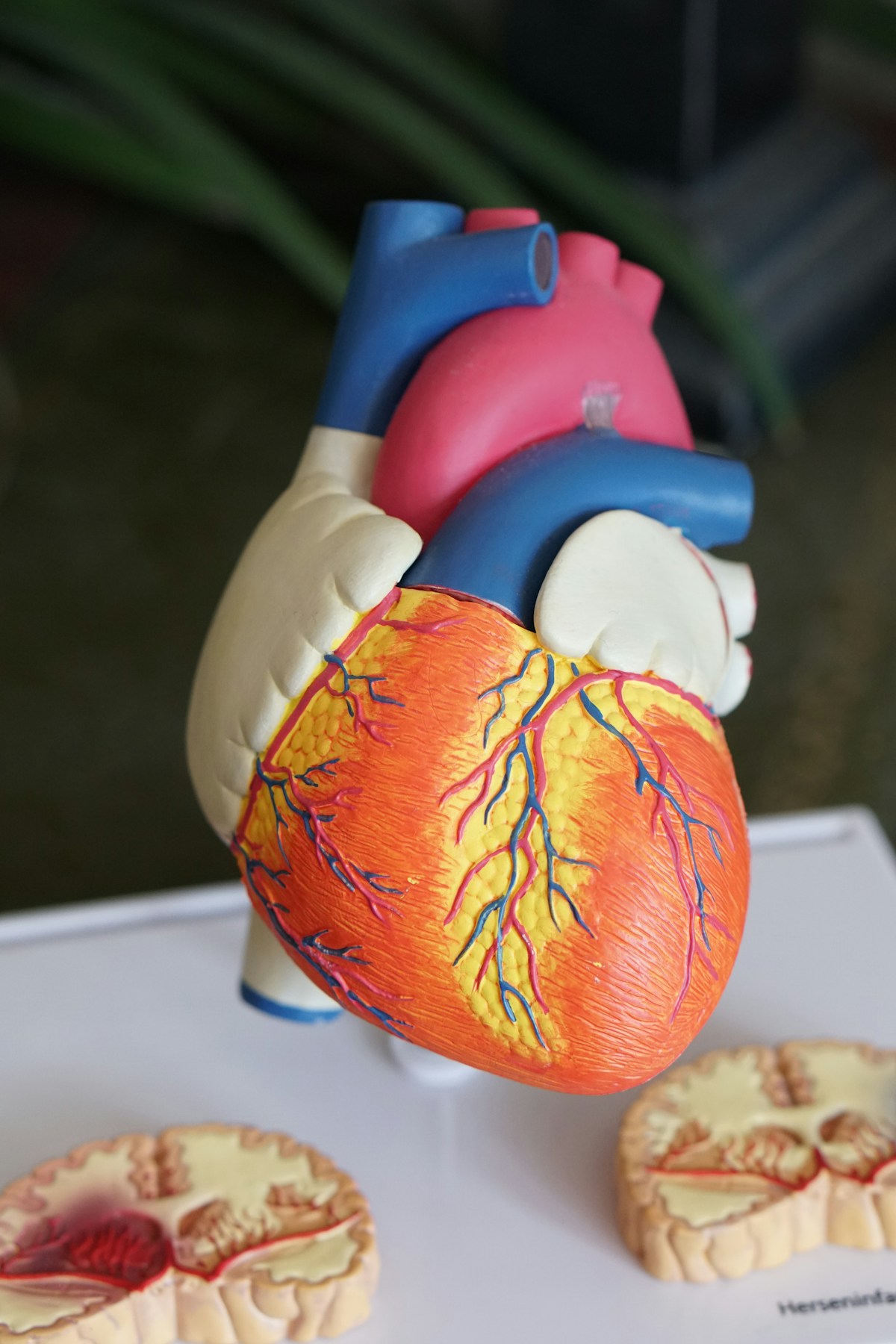
Understanding Disc Anatomy
The intervertebral disc has two main components:
The nucleus pulposus — the inner gel-like core, composed largely of water-binding proteoglycans. This is the high-pressure, shock-absorbing component of the disc.
The annulus fibrosus — the outer fibrous ring, composed of multiple layers of collagen fibers arranged at alternating angles. This provides tensile strength and contains the nucleus.
Disc herniation occurs when the nucleus breaks through tears in the annulus and either bulges against the annular outer fibers (protrusion) or completely escapes through a full-thickness annular tear (extrusion or sequestration). The extruded material can then compress adjacent nerve roots.
The annular tears that allow herniation develop gradually under repeated abnormal mechanical loading — they don't occur from a single incident in most cases. The "injury" that seems to cause a disc herniation (a lift, a twist, a sneeze) is usually the final straw applied to a disc that has been progressively weakened by years of abnormal loading.
Why Standard Treatments Are Incomplete
Bed Rest and Pain Medication
These approaches address the symptom but not the structural cause. They are appropriate as short-term management while the acute nerve irritation settles, but they do nothing to change the mechanical environment that caused the herniation.
Epidural Steroid Injections
ESIs reduce the inflammatory component of nerve root irritation and can provide meaningful temporary relief. Multiple studies show they are effective for short-term symptom reduction. However, they do not change disc pathology, they do not improve long-term outcomes compared to no injection, and they do nothing to address the structural mechanics that drove the herniation.
Surgery (Microdiscectomy)
Surgical removal of the herniated disc material — microdiscectomy — is effective and appropriate in specific circumstances: severe or progressive neurological compromise, cauda equina syndrome, or failed conservative care over an adequate period. When indicated, it can provide faster relief than conservative care. But several important considerations are consistently overlooked:
- Adjacent segment degeneration: Operated discs have altered biomechanics that increase stress on adjacent levels, accelerating degeneration above or below the surgical site.
- Recurrence: Disc herniation recurs at the same level in approximately 5–10% of cases, and adjacent level problems develop in a significant proportion over 5–10 years.
- The structural problem remains: Surgery removes the herniated material but doesn't correct the loading pattern that caused the herniation. Without structural correction, the post-surgical spine continues to be mechanically vulnerable.

The Structural Framework for Disc Care
A comprehensive structural approach to disc herniation encompasses several dimensions that standard care ignores:
Restoring lumbar lordosis
The lumbar lordosis — the natural inward curve of the lower back — is mechanically protective for the discs. A properly curved lumbar spine loads disc surfaces evenly, reduces posterior disc stress, and maintains appropriate foraminal dimensions. Loss of lordosis (which is ubiquitous in desk workers and chronic sitters) concentrates compressive stress at the posterior disc margin — exactly where herniations occur.
Structural protocols specifically aimed at restoring lumbar lordosis — through traction, specific mobilization, and progressive extension loading — can measurably change disc loading and are supported by evidence.
Pelvic Alignment Correction
An unlevel pelvis or sacral base obliquity creates asymmetric loading across the disc surface. If the pelvis is higher on the right, the L5-S1 disc is loaded more heavily on the right side — which helps explain why disc herniations tend to be unilateral and why they occur at specific levels rather than randomly.
Correcting pelvic alignment doesn't just help symptoms — it changes the mechanical environment of the disc.
Decompression
Specific traction techniques that reduce the compressive load on herniated discs can facilitate the natural resorption process. By creating intermittent negative pressure within the disc, traction may draw disc material back toward the nucleus and reduce the size of the protrusion over time.
Neuromuscular Stabilization
The multifidus muscle — the deep spinal stabilizer at each vertebral level — undergoes rapid atrophy after disc injury. This isn't a voluntary process; it is a neurological inhibition response. Rebuilding multifidus activation and endurance is essential for preventing recurrence after disc herniation.
The Natural Healing Process
There is genuinely good news about disc herniation that most patients are never told: the immune system can and does resorb herniated disc material. Studies using serial MRI show that large disc herniations (particularly extrusions and sequestrations) often significantly reduce in size over 6–24 months. The extruded disc material is recognized as foreign by the body's immune system and gradually removed.
This process is not inevitable and not uniform — some herniations resorb completely, others partially, others not at all. But it is common enough to be the standard expectation for large herniations managed conservatively. Structural care during this period supports the process by reducing mechanical stress on the healing tissue.
Frequently Asked Questions
Q: My disc herniation is "moderate to large" — should I be considering surgery?
Size alone is not an indication for surgery. The primary indications are: significant and progressive neurological deficit (especially leg weakness), cauda equina syndrome (bladder/bowel involvement), or failure of an adequate course of quality conservative care. Many large herniations resolve without surgery. An evaluation will determine whether your specific situation has features that suggest conservative or surgical management.
Q: How long does disc herniation take to heal?
With appropriate structural management, most people with acute disc herniation experience meaningful improvement within 6–12 weeks. Complete resolution of structural findings on MRI (if they resorb) typically takes several months. Residual imaging findings after symptom resolution are common and are not clinically significant.
Q: Can I still exercise with a herniated disc?
With guidance, yes. The key is identifying which movements and loads are appropriate for your specific herniation pattern. Extension-biased loading (which increases lumbar lordosis) is often well-tolerated and beneficial for posterior disc herniations. Flexion-biased loading (forward bending, sit-ups) is typically contraindicated initially. A specific exercise program tailored to your structural findings is far more valuable than generic advice.
Q: Will I always have a "bad back" after a disc herniation?
Not necessarily. People who receive appropriate structural care, address the underlying mechanics that drove the herniation, and rebuild proper neuromuscular stabilization can achieve a fully functional, pain-free back. The structural correction phase takes months, but the long-term outcome is a structurally sounder spine than they had before — because the underlying problems that caused the herniation have been addressed.
Conclusion
Your MRI is a piece of the puzzle — an important one, but only one. A herniated disc is not a disease; it is a structural consequence of years of abnormal loading. Treating it without understanding and correcting those loading patterns leaves the underlying problem in place and makes recurrence predictable.
At SPINE-X, we interpret your imaging in the context of a full structural evaluation — understanding not just what your disc looks like, but why it herniated, what structural corrections are needed, and how to rebuild the mechanical foundation that will keep it from happening again.
Related Reading
- Sciatica: Why It's Not What Most People Think
- Can a Herniated Disc Heal on Its Own? The Evidence Explained
- The SPINE-X Approach to Herniated Disc: Conservative Care That Actually Works
Is Your Spine Contributing to Your Symptoms?
Reading about structural problems is one thing — knowing what is actually happening in your spine is another.
Dr. Joy offers a personal Diagnostic Report — send 4 posture photos, and receive a detailed written analysis of your structural findings, postural deviations, and a personalized exercise and correction plan. All delivered as a PDF within 48 hours.
- Postural deviation analysis (anterior, posterior, lateral views)
- Structural findings: curvature, head position, pelvic levelness
- Personalized correction and exercise recommendations
- PDF report you can reference at home
$40 · Remote · Results in 48 hours
Reviewed by Dr. Ji Young Lim, D.C. — 13+ years clinical experience in structural chiropractic